Endoscopes and Infection: Design Matters

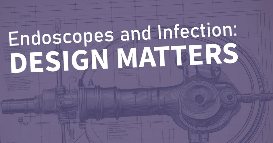
 There are four fundamental aspects of endoscopes that lead to infection: Intricate design, biofilm formation, human error during reprocessing, and failure to dry properly. Much emphasis is being placed on the first: The intricate design of this technology that provides so many reservoirs for bacteria to escape cleaning and be transmitted to a vulnerable host. In today's post, we'll look at how design improvements have led to safer endoscopes, and what we can look forward to in the future of endoscope design.
First, let's go back to the first endoscope. In 1868, German physician Adolph Kussmaul designed a way to visualize the stomach via a tube inserted into the mouth. But who could tolerate such discomfort long enough for him to try it out? Well, a professional sword-swallower, of course! Having procured his sword-swallower from a local circus, Dr. Kussmaul proceeded to test out his theory - only to discover that the interior of the body is far too dark to visualize anything at all. However, Dr. Kussmaul's first step led to further advancements over the next 100 years: Use of electric light, bendable tubing, fiber optics, digital cameras, and even biopsy tools. The result is a highly complex, versatile technology that is essential for modern visualization, diagnosis, and even treatment of a huge range of illnesses. But with this complexity came the intricate designs that can lead to bioburden buildup and infection outbreaks.
Now let's consider what Dr. Kussmaul's invention looks like today. Endoscopes, regardless of type, are made up of three main components. The endoscope, the main body (video monitor), and peripherals.![]()
The endoscope itself has three parts as well. The control section features all the knobs necessary to control the camera, lights, and attachments as well as to maneuver the device safely in the patient's body. On one end, the control section is connected via the universal cord to the main body (video monitor) and other related peripherals that provide the endoscope with power, suction, and water. Connected to the other side of the control section is the insertion tube, the flexible tube that enters the patient's body. This half-inch or narrower tube contains bundled channels for water, suction, device controllers, imaging, and lighting. The tip of the tube contains sealed lenses and lights, but also an open channel where device attachments are placed. This channel has been implicated in outbreaks, and presents the greatest threat for biofilms.
So what is being done to make endoscopes safer?
- Badly designed endoscopes have been recalled. This applies to endoscopes whose moving parts were shown to resist even the most meticulous cleaning.
- New designs allow impossible-to-clean components to be single-use or removable for cleaning. This allows the benefits of the complex technology without the contamination risk.
- Single-use endoscopes are available for the most vulnerable patients. While this costly option is not sustainable for all patients, those for whom an infection would cause the greatest harm benefit from a single-use instrument.
- Automatic reprocessors add an extra layer of safety, since they have timers and sensors to make sure all steps are completed.
- Technologies in reprocessing, such as surveillance cameras that go into the device, as well as redesigned drying cabinets provide additional safety nets.
- New innovations, such as swallowable camera capsules, may provide new options for visualization.
Looking to the future, exciting ideas are being investigated. Remote-control visualizers could make insertion tubes less complex and thinner, leading to more comfort and less surface area. New materials that prevent biofilms could help prevent bioburden contamination, while new designs could allow for more access for disinfection.
What changes in endoscopes have you seen in your facility? Share your experiences in the comments below!
![EOScu Logo - Dark - Outlined [07182023]-01](https://blog.eoscu.com/hubfs/Eoscu_June2024/Images/EOScu%20Logo%20-%20Dark%20-%20Outlined%20%5B07182023%5D-01.svg)


